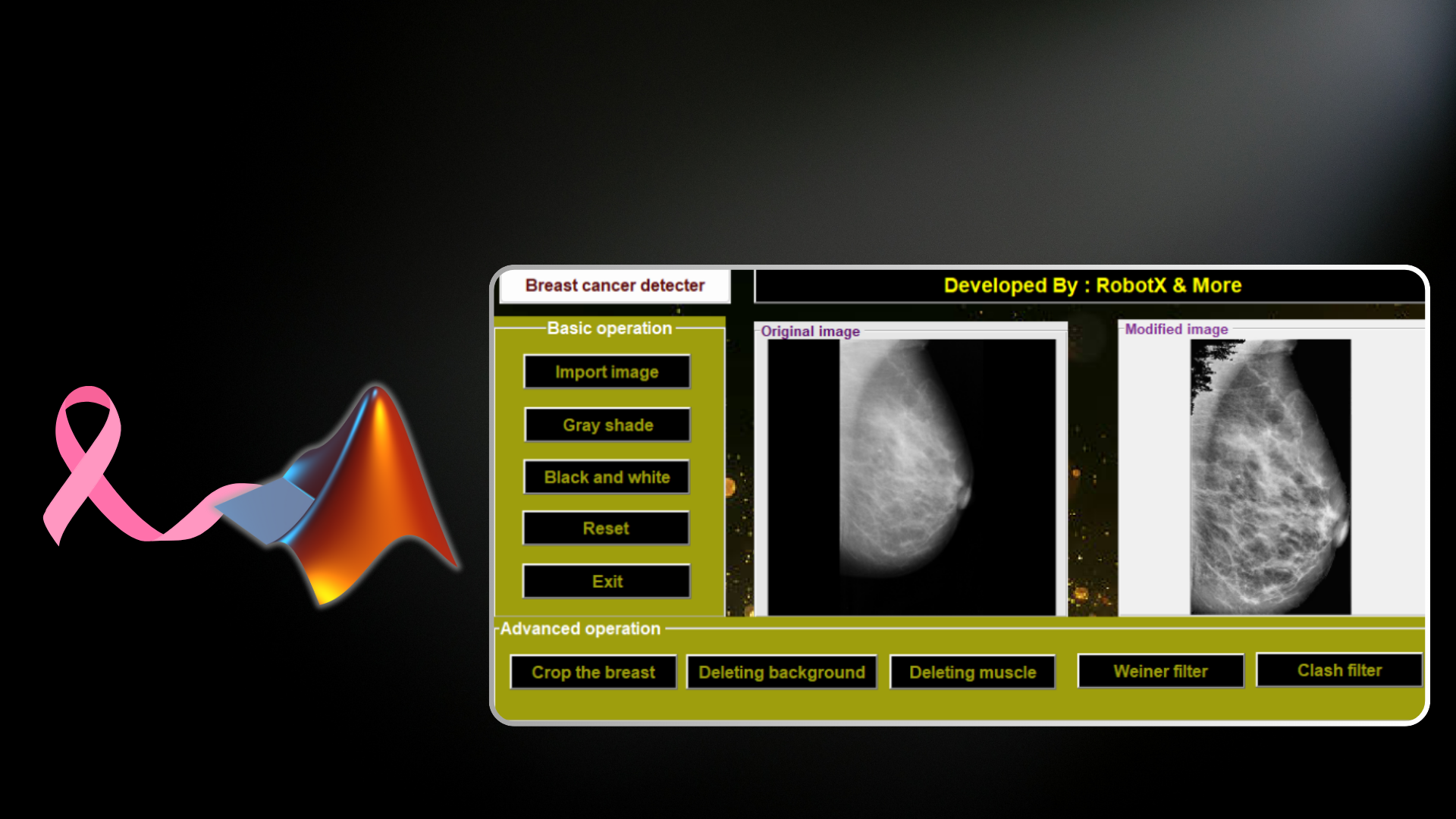
 GitHub Repository
Overview
This project addresses one of the most critical challenges in medical imaging: enhancing mammography images for improved breast cancer detection. Early detection of breast cancer significantly increases survival rates, but standard mammography images often contain artifacts and noise that complicate diagnosis. Our application implements a comprehensive set of image pre-processing techniques to remove unwanted artifacts, enhance image quality, and extract precise regions of interest (ROI), thereby assisting radiologists in making faster and more accurate diagnoses.
Problem Statement
Mammography images frequently contain several challenges that hinder accurate diagnosis:
- Background noise and artifacts
- Presence of pectoral muscle that can be mistaken for abnormalities
- Poor contrast and image quality
- Difficulty in identifying the exact region of interest
These issues can lead to missed diagnoses or false positives, both of which have serious implications for patient care.
Solution Architecture
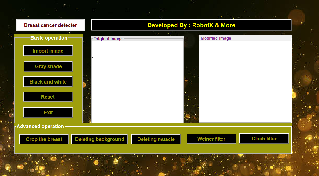
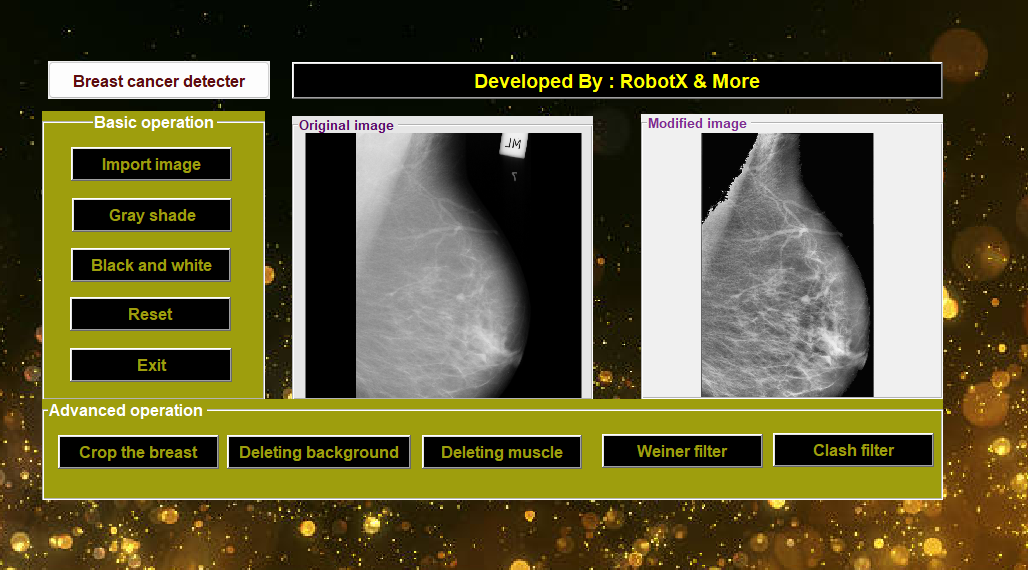
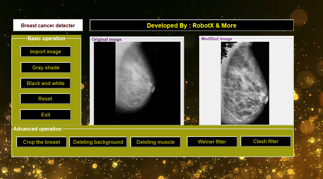
We developed a MATLAB-based application with a user-friendly GUI that allows medical professionals to:
- Import mammography images
- Apply various image processing techniques
- View the original and enhanced images side-by-side
- Save processed images for further analysis
Key Features and Technologies
Basic Operations
- Image Import: Supports standard medical imaging formats
- Grayscale Conversion: Transforms colored images to grayscale for standardization
- Black and White Transformation: Applies binary thresholding for enhanced feature distinction
- Reset and Exit Functionality: Ensures user-friendly experience
Advanced Operations
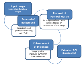
- Breast Region Extraction: Isolates the breast tissue from the background
- Background Removal: Eliminates unwanted artifacts using thresholding techniques
- Pectoral Muscle Removal: Implements modified region growing algorithm to remove the pectoral muscle
- Wiener Filter: Reduces noise while preserving edges and important features
- CLAHE (Contrast Limited Adaptive Histogram Equalization): Enhances image contrast for better visualization of abnormalities
Technical Implementation
The application was built using:
- MATLAB R2020a: Primary development environment
- Image Processing Toolbox: For specialized image manipulation algorithms
- GUIDE (GUI Development Environment): For creating the user interface
- mini-MIAS Database: For testing and validation (Mammographic Image Analysis Society database)
Advanced Algorithms Implemented:
- Thresholding with Connected Component Analysis: For background removal
- Modified Region Growing Technique: For pectoral muscle suppression
- Automatic seed point selection based on image orientation
- Adaptive region growing based on pixel intensity
- Optimized Wiener Filter: Applied with a [3,3] filter mask for optimal noise reduction
- CLAHE with Contrast Index 0.2: For optimal contrast enhancement
Results and Validation
The application was tested on 100 images from the mini-MIAS database, resulting in:
- 98% Correctness Rate: Accurate segmentation and artifact removal
- 97% Completeness Rate: Successful ROI extraction
- Significant Improvement in Image Quality: Measured by PSNR (Peak Signal-to-Noise Ratio) and IQI (Image Quality Index)
- Enhanced Visualization: Better contrast and clarity for identifying potential abnormalities
The performance metrics showed that the optimized parameters ([3,3] filter mask for Wiener filter and 0.2 Contrast Index for CLAHE) provided the best balance between noise reduction and feature preservation.


Impact and Applications
This project has several practical applications in medical diagnosis:
- Assists Radiologists: Helps medical professionals identify subtle changes in breast tissue
- Reduces Diagnostic Time: Streamlines the image analysis process
- Improves Early Detection: Enhances the visibility of small abnormalities
- Educational Tool: Can be used for training medical students and residents
- Research Platform: Provides a foundation for further development of CAD (Computer-Aided Diagnosis) systems
Future Enhancements
- Implementation of machine learning algorithms for automatic abnormality detection
- Integration with hospital PACS (Picture Archiving and Communication System)
- Development of a cloud-based version for remote diagnosis
- Extension to other medical imaging modalities beyond mammography
Contributors
- Imad-Eddine NACIRI
- Achraf Berriane
- Errouji Oussama
